Since its establishment, the Department of Ophthalmology at Taiwan Adventist Hospital has been committed to providing patients with professional consultations and comprehensive medical care. We continuously bolster our team of specialized physicians and continually introduce the latest technologies and equipment to ensure precise diagnosis and treatment.
Our Services
Service and Treatment
Examination Items
Management & Treatments
Health Education Information
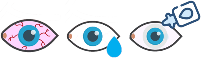
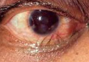
Dry eye disease is a prevalent condition that occurs when the eyes don't produce enough tears or when the tears evaporate rapidly. This issue has become particularly widespread among modern individuals who often strain their eyes by prolonged use of computer screens without breaks, leading to a decrease in the age of affected patients.
The Department is equipped with professional examination equipment providing comprehensive treatment and follow-up care for dry eye patients.
Symptoms & Causes
■ Symptoms:
itching
eyelids stuck together upon waking
crusty debris around the eye lashes
eyelid red
burning
granular sensation and scratchiness or foreign-body sensation due to crusted debris or dryness
decreased vision or changes in visual clarity due to poor tear film
sensitive to light
excessive tearing
■ Dry eyes have many causes, such as:
over the age of 50
wearing contact lenses
looking at computer screens for a long time without a break
Spending time in air conditioned, heated, windy, cold, dry or dusty environments
drug-induced (for example, some antidepressants or blood pressure medicines)
other systemic disease related, such as blepharitis, Sjögren's syndrome or lupus and more
Many people get dry eyes, which is not usually serious and there are things you can do to help. Dry eye is a chronic disease; it requires long-term treatment and care, which can often be managed, but not completely cured.
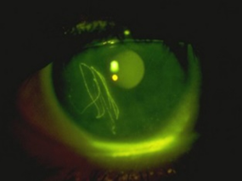
Dry eye syndrome is mainly divided into oil-deficient type and water-deficient type. Examination can help doctors determine the main factors causing dry eye, and facilitate the selection of subsequent treatment strategies.
Tests and procedures that may be used to determine the cause of your dry eyes include:
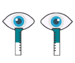
Schirmer’s Test: A test to measure the volume of your tears. In this test, blotting strips of paper are placed under your lower eyelids. After five minutes, the ophthalmologist measures the amount of strip soaked by your tears.
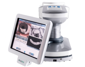
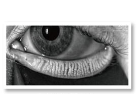
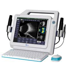
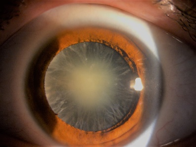
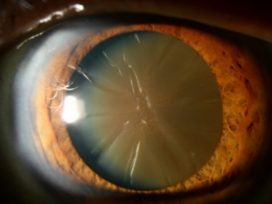
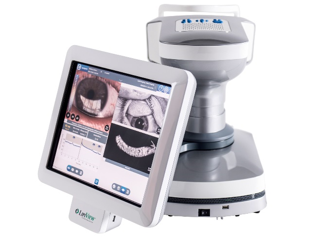
LipiView Interferometer: It helps the physicians to measure the lipid layer thickness, observe the dynamic Meibomian gland and evaluate patient’s blinking function.
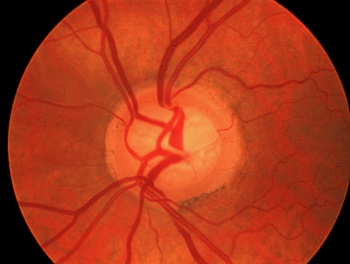
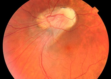
Normal Meibomian Gland Structure
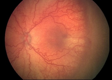
Meibomian Gland Atrophy or Loss
When Meibomian gland blockage is not ruled out, it will easily lead to:
Meibomian degeneration.
Meibomian atrophy.
Uneven secretion of the lipid layer of the tear film.
Unable to protect the water layer, that causes the watery tears to dry up more quickly and easily (no matter how much artificial tears have applied).
Treatments for dry eyes may make you more comfortable. These treatments include lifestyle changes and eye drops. You'll likely need to take these measures indefinitely to control the symptoms.
Daily Care for Eyes: Take breaks to rest your eyes, maintain eye hygiene routine, and apply warm compress around the eyes habitually.
Medical Treatment: After being diagnosed by the physician, use artificial tears or gel to keep the surface of the eyes moist.
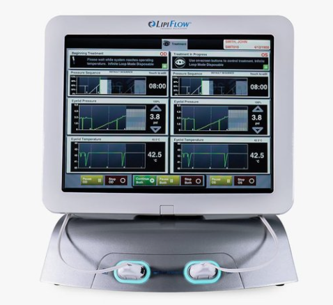
LipiFlow: Lipiflow is a ground-breaking technology, which is a heat treatment for Meibomian gland dysfunction. It helps to remove these blockages in a warm, soothing way, then allows the body to resume the natural production of lipids (oils) needed for the tear film.
Common eye diseases and cases where individuals experience discomfort but are unable to determine the specific condition. Examples include conjunctivitis, driver's license eye examinations, basic eye examinations, and refraction tests for eyeglasses fitting, among others.
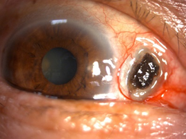
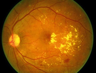
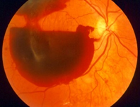
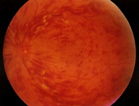
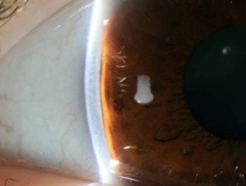
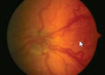
The Retina Clinic primarily operates as an outpatient facility, specializing in comprehensive eye examinations and investigations pertaining to conditions such as floaters, vitreous/retinal hemorrhage, breaks or detachments, macular degeneration, retinal inflammatory dystrophy, uveitis, retinal pathologies associated with diabetes and hypertension, high myopia retinal examination, and more.
Our team of esteemed physicians possesses profound expertise and extensive research acumen in the realm of retinal-related diseases. Coupled with cutting-edge diagnostic and surgical equipment, we deliver a holistic spectrum of treatments and meticulous follow-up care tailored to patients afflicted with dry eye syndrome.
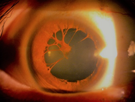
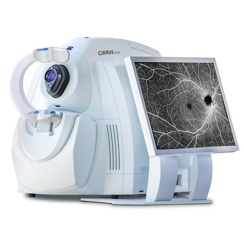
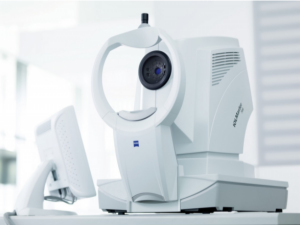
Zeiss Cirrus Angio OCT 6000/600 - Retinal Vascular Optical Coherence Tomography Scanner Zeiss Cirrus Angio OCT 6000/600 provides high-resolution cross-sectional or longitudinal profiles of various parts of the eye, ranging from the retina to the choroid. This advanced imaging technology enables a physician understanding of the location and nature of pathological conditions, thereby facilitating diagnosis and treatment.
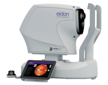
Eidon - True Color Confocal Fundus Scanner The scanner allows for a quick assessment of the entire condition of the fundus without the need for pupil dilation. It provides comprehensive fundus retinal images for medical professionals to analyze, enabling the early detection of pathological changes and achieving preventive and therapeutic effects.
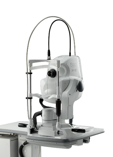
NIDEK Mirante SLO/FAG/ICG When combined with contrast agents, it allows for the capture of images of retinal blood vessels. This aids in the diagnosis and monitoring of retinal/choroidal-related diseases, such as retinal neovascularization, retinal hemorrhage, and vessel
occlusion, among others.
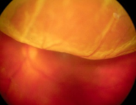
MEDA A/B Scan MD-2300S Uses ultrasound to scan ocular cross-sectional images, aiding doctors in diagnosing eye conditions such as vitreous hemorrhage and retinal detachment, among others.
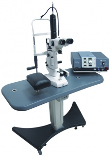
Argon Laser treatment is used to coagulate the peripheral retina, with the aim of preventing the formation of new blood vessels and reducing the risk of vitreous hemorrhage and retinal detachment, which can lead to vision impairment.
Pars Plana Vitrectomy (PPV) is a surgical procedure in which pathological vitreous humor is removed from the eye using surgical instruments. Depending on the situation, other substances such as gas or silicone oil may be injected.
Service and Treatment
Examination Items
Glaucoma is a general term used to describe a group of eye disorders that damage the optic nerve, which is responsible for transmitting visual information to the brain. It is the most common cause of optic nerve damage leading to vision loss. Regardless of the specific type of glaucoma, individuals may encounter the following symptoms:
Eye pain or pressure.
Headaches.
Seeing rainbow-colored halos around lights.
Experiencing low vision, blurred vision, narrowed vision (known as tunnel vision), or blind spots.
Nausea and vomiting.
Redness in the eyes.
Our team of medical professionals boasts a wealth of clinical experience, augmented by our department's state-of-the-art glaucoma-specific diagnostic equipment. This advanced technological infrastructure empowers us to deliver thorough monitoring and comprehensive treatment for individuals afflicted by glaucoma.
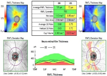
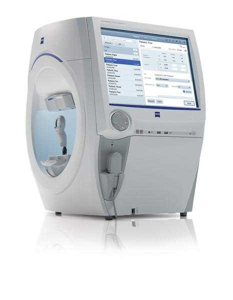
The Zeiss Cirrus Angio OCT 6000/600 can provide high-resolution cross-sectional or longitudinal profiles of the optic nerve, enabling physicians to gain a better understanding of the location of pathological conditions and aiding in diagnosis and treatment.
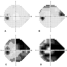
The Humphrey Field Analyzer 3 is used to examine for the presence of visual field defects, enabling early diagnosis of conditions such as glaucoma, optic nerve issues, and brain abnormalities.
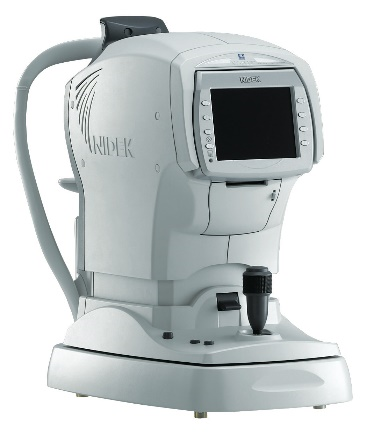
The NIDEK NT-530P The NIDEK NT-530P is used to measure intraocular pressure and corneal thickness, checking for elevated eye pressure due to thicker corneas.
Service and Treatment
Examination Items
Management & Treatments
Health Education Information
Cataracts can lead to blurred vision and heightened sensitivity to glare from lights. If a cataract hinders your ability to carry out everyday activities, your physician may recommend cataract surgery.
Furthermore, when a cataract complicates the management of another ocular condition, cataract surgery might be advised. For instance, if a cataract obstructs your eye doctor's ability to examine the posterior segment of your eye for the monitoring or treatment of conditions such as age-related macular degeneration or diabetic retinopathy, cataract surgery may be proposed.
Cataract surgery is a specialized procedure designed to remove the eye's lens and, in most scenarios, replace it with an artificial lens. Ordinarily, the eye's lens is transparent. However, the presence of a cataract leads to clouding, which eventually affects vision.
This surgery is performed on an outpatient basis, meaning there's no need for a post-surgery hospital stay. Cataract surgery is a routine and generally safe procedure, widely practiced within the medical field.
Our medical team possesses extensive clinical diagnostic and surgical experience, providing patients with comprehensive follow-up care. Additionally, our department is equipped with cutting-edge phacoemulsification surgical instruments for cataract treatment. This advanced technology not only enhances surgical outcomes but also reduces the risk of postoperative infections, while minimizing incision size.
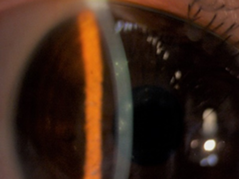
The ZEISS IOL-MASTER 700, a non-contact precision artificial intraocular lens measurement device, is used for pre-cataract surgery examinations. It precisely measures corneal curvature, axial length, and anterior chamber depth to calculate the required power for artificial intraocular lenses.
The MEDA A/B Scan MD-2300S, A-scan ultrasound provides measurements of corneal thickness, lens thickness, axial length, and anterior chamber depth to calculate the power of artificial intraocular lenses.
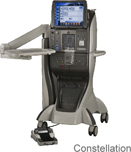
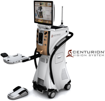
Cataract Ultrasonic Phacoemulsification Surgery with CENTURION® The most advanced cataract ultrasonic phacoemulsification device, offering minimally invasive, sutureless surgery.
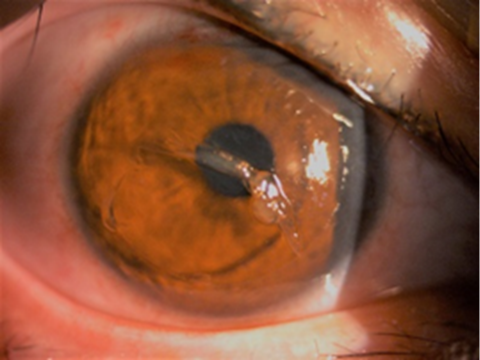
LightLas YAG Laser Photodisruptor Lightmed, Taiwan Lightmed YAG Laser Photodisruptor for Ophthalmology. Laser treatment for secondary cataracts.
Pediatric Vision Care Clinic incorporate special techniques and technology to ensure accurate testing and help to accommodating and monitoring children's visual health, conducting examinations for amblyopia (lazy eye) and strabismus, addressing refractive errors, and fitting (contact) lenses. Additionally, we collaborate with school vision screenings and provide education on vision protection and prevention, ensuring comprehensive eye care for children.
The Eye Exam
In addition to basic visual acuity (distance and near vision, or refractive errors) an eye exam may assess the following visual skills that are required for learning and mobility:
Binocular vision: how the eyes work together as a team
Focusing
Peripheral Vision
Color Vision
Hand-eye Coordination
Tracking
Stereopsis
The doctor will also examine the area around the eye and inside the eye to check for any eye diseases or health conditions. You should tell the doctor any relevant personal history of your child such as a premature birth, developmental delays, family history of eye problems, eye injuries or medications the child is taking. This would also be the time to address any concerns or issues your child has that might indicate a vision problem.
Children’s Eyeglasses, Contacts & Other Treatments
If the eye doctor does determine that your child has a vision problem, they may discuss a number of therapeutic options such as eyeglasses or contact lenses, an eye patch, vision therapy or orthokeratoplasty, depending on the condition and the doctor’s specialty. Since some conditions are much easier to treat when they are caught early while the eyes are still developing, it is important to diagnose any eye and vision issues as early as possible.
We provide professional consultation and treatment for conditions such as eyelid turning in(entropion)/out(ectropion), nasolacrimal duct blockage, excessive tearing, inverted eyelashes, and eyelid plastic surgery.
In collaboration with the Neonatology department, our clinic offers comprehensive follow-up examinations and treatments to monitor and address the ocular and retinal development of newborns and premature infants.
Tracks changes in visual fields caused by glaucoma.
Examines visual field defects caused by brain and other factors.
Conducts international standard visual field tests for driver's licenses.
Eidon - True Color Confocal Fundus Scanner
Utilizing confocal scanning with different light sources, including white light LED (440-650 nm) and near-infrared LED (825-870 nm), for imaging, creating three-dimensional and true-to-life images.
Single images cover up to 60°, and multiple images can be stitched together to achieve a wide-angle view of up to 110°.
Retinal images can be captured without the need for pupil dilation, with a minimum pupil size of 2 mm.
Assisting in the diagnosis of various retinal diseases, tracking changes in diabetic retinopathy, and monitoring glaucoma-related optic nerve conditions.
Precise measurement of corneal curvature, axial length, and anterior chamber depth for pre-cataract surgery examinations, enabling accurate calculation of artificial intraocular lens power.
Utilizes SWEPT Source OCT technology for faster optical measurements and higher cataract penetration rates.
Incorporates the latest Barrett formulas to provide more precise calculations of intraocular lens power.
LipiView - Dual Digital Meibomian Gland Imaging Device
Detects tear film (lipid layer) thickness, meibomian gland health, and checks if blinking function is normal.
Provides physicians with insights into the primary causes of dry eye syndrome, aiding in treatment decisions.
LipiFlow - Thermal Pulsation Treatment Device
FDA-certified treatment for meibomian gland dysfunction.
12 minutes of pulsating warmth at 42.5°C.
Effectively softens obstructed glands, clears meibomian glands, and promotes the secretion of new lipids, preventing gland atrophy caused by blockages.
Non-contact examination with automatic focusing, measurement, and automatic analysis of endothelial cells (16 images per second, rapid analysis in 2 seconds).
Evaluates corneal endothelial cell density, morphology, hexagonal cell count, and coefficient of variation.
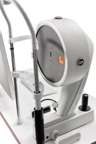
Pentacam HR 70900 - Corneal Imaging Analyzer
Rapidly scans 100 images from multiple angles for comprehensive corneal assessments.
Provides high-resolution corneal layers and density for the diagnosis of corneal diseases.
Assists in interpreting data related to glaucoma.
Early diagnosis of keratoconus for pre/post-cataract surgery examinations.
 Normal Meibomian Gland Structure
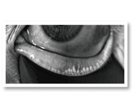 Meibomian Gland Atrophy or Loss


 黃峰霖 醫師
 林于皓 醫師